THE LACRIMAL BONE
The Lacrimal Bone: Ever wondered how you tear up and what is involved in the formation of tears? Well, in this article, you will uncover the lacrimal bone and its involvement in spurting teardrops from your eyes.
What Is The Lacrimal Bone?
The lacrimal bone is one of the bones that make up the skull. It is the smallest and most brittle bone of the face and is located in front of the inner wall of the eye socket.
The lacrimal bone has two sides and four edges. It is square and is located in front of the inner wall of the binocular kiln. A thin bone in front of the medial wall of the orbit, with a groove in which the lacrimal sac fits.
It is a pair of left and right irregular rectangular thin bones that form the latter half of the fossa for the lacrimal sac at the anterior end of the sidewall of the orbit, which also forms part of the bone wall of the nasolacrimal duct that follows. This bone is also produced by connective tissue ossification.

The overall shape of the lacrimal bone resembles a fingernail but is thinner than the nail. The outer surface faces the orbit, and the groove-shaped depression in front of the longitudinal ridge is the part that adds to the composition of the fossa for the lacrimal sac.
The outer surface forms the anterior part of the inner wall of the orbit, and the inner surface forms part of the outer wall of the nasal cavity (middle nasal meatus). The upper edge is in contact with the frontal orbit, the lower edge is in contact with the maxillary orbital surface, the anterior edge is in contact with the maxillary frontal process, and the posterior edge is in contact with the ethmoid orbital plate.
In the anterior half of the lateral surface is a longitudinal lacrimal groove that, together with the same-named groove in the frontal process of the maxilla, forms the fossa for lacrimal.
The posterior boundary of the lacrimal sac is called the posterior lacrimal ridge, which extends downward to form the lacrimal groove and forms part of the nasolacrimal duct wall together with the lacrimal groove of the frontal process of the maxillary bone and the lacrimal process of the inferior turbinate. do. Lacrimale is an adjective for lacrimal (tears).
Where Is The Lacrimal Bone Located?
The lacrimal is located in the anterior part of the medial wall of the orbit. It has two surfaces and four boundaries. Several bony landmarks of the lacrimal bone function in the process of lacrimation or weeping. Specifically, the lacrimal bone helps to form the nasolacrimal canal which is needed for the translocation of tears.
A depression on the anterior inferior part of the bone, the lacrimal fossa contains a membranous lacrimal sac. Tears or lacrimal fluid from the lacrimal glands are stored in this sac in cases of excessive lacrimation. The fluid then passes through the nasolacrimal duct and into the nasopharynx. This drainage usually results in a runny nose during excessive crying or tear production. If the lacrimal bone is injured or fractured, a posttraumatic blockage of the lacrimal pathway can arise.
SEE RELATED POST: All You Need To Know Tortuous Colon
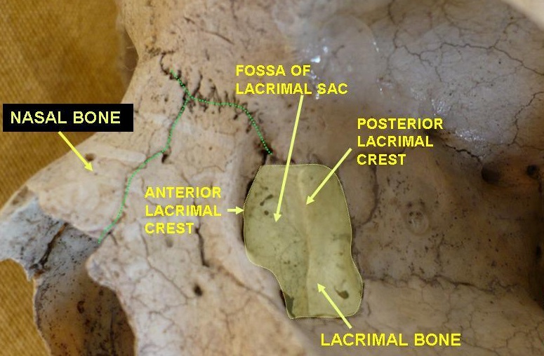
The lateral or orbital surface is divided into two by a vertical ridge, the posterior lacrimal crest. In front of this crest is a longitudinal groove, the inner margin of the lacrimal sulcus (sulcus lacrimalis), which unites with the frontal process of the maxilla, and the lacrimal fossa is thus complete. The upper part of this fossa contains the lacrimal sac, the lower part, the nasolacrimal duct.
/Lacrimal-b0855fbf3b6047aca80784c46a9c2e5f.jpg)
The posterior part of the crest is smooth and forms part of the medial wall of the orbit. The crest, with a part of the orbital surface immediately behind it, attaches to the lacrimal part of the origin orbicularis oculi and ends below in a small, hook-like projection, the lacrimal Humulus, which articulates with the lacrimal tubercle of the maxilla, and completes the upper opening of the nasolacrimal canal; the hamulus is sometimes present as a separate fragment and is then called the lesser lacrimal bone.
The Lacrimal Bone: Medial And Nasal Surface
The medial or nasal surface presents a longitudinal groove corresponding to the crest on the lateral surface. The area in front of this groove becomes part of the middle of the nose. Its posterior region articulates with the ethmoid and meets some of the anterior ethmoidal cells.
Borders Of the four limits: articulates with the frontal process of the anterior maxilla; lamellae along the back of the papyracea trellis; With the superior frontal bone. The interior is divided by the lower end of the posterior lacrimal crest into two parts:
The posterior part is associated with the orbital plate of the maxilla;
The anterior descends as a descending process, articulates with the lacrimal process of the inferior nasal concha, and helps form the canal for the nasolacrimal duct.
Development
The lacrimal fracture occurs from a single-center, which appears about week twelfth in the membrane covering the cartilaginous nasal capsule.
Joint
The lacrimal bone articulates with four bones: two cranial, frontal and ethmoid, and two facial, maxilla, and inferior nasal concha.
Other Animals
In early lobe-finned fishes and ancestral tetrapods, the lacrimal bone is a relatively large and strong bone, running from the orbit to the nostrils. It forms the side of the face, occurring in the middle of the nasal bones and the maxilla. In primitive forms, it is often accompanied by a much smaller septomaxilla bone, which is immediately behind the nasal opening, but it is lost in most modern species.
The lacrimal bone is often smaller in living vertebrates and is no longer always directly attached to the nasal opening, although it retains its connection with the orbit. The lacrimal bone is completely absent in living amphibians, as well as in some reptile species.
If you found this article educating, kindly share and comment. Thanks for reading!
Sharing Is Caring!